
Equipment is only useful if it changes the decision.
We don't buy technology to have it on a list. Each of these changes something concrete — what we can see before we operate, how much of the surgery we can plan in advance, or how much you have to endure.
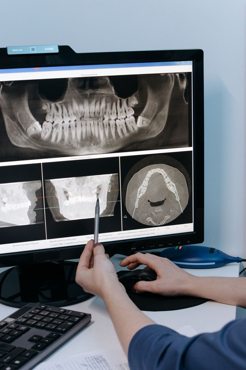
CBCT 3D imaging
A standard dental x-ray is flat. It shows you a shadow, and it cannot tell you how wide a piece of bone is, or exactly where a nerve runs, or how far a sinus has dropped.
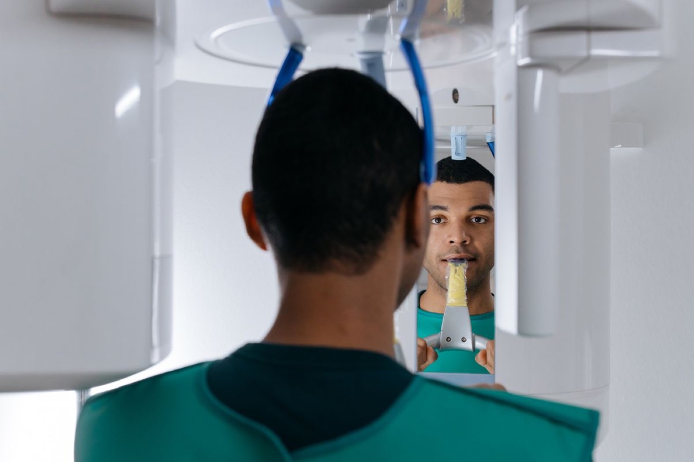
A CBCT scan is a three-dimensional image of your jaw. We can measure bone in every direction, see the nerve canal precisely, and place each implant virtually — choosing the angle, depth and size — before we place it in you.
It is also the reason some patients who were told elsewhere that implants were impossible turn out to be perfectly good candidates. Flat x-rays hide bone that a 3D scan finds. The scan takes under a minute and uses a low radiation dose.
LANAP laser
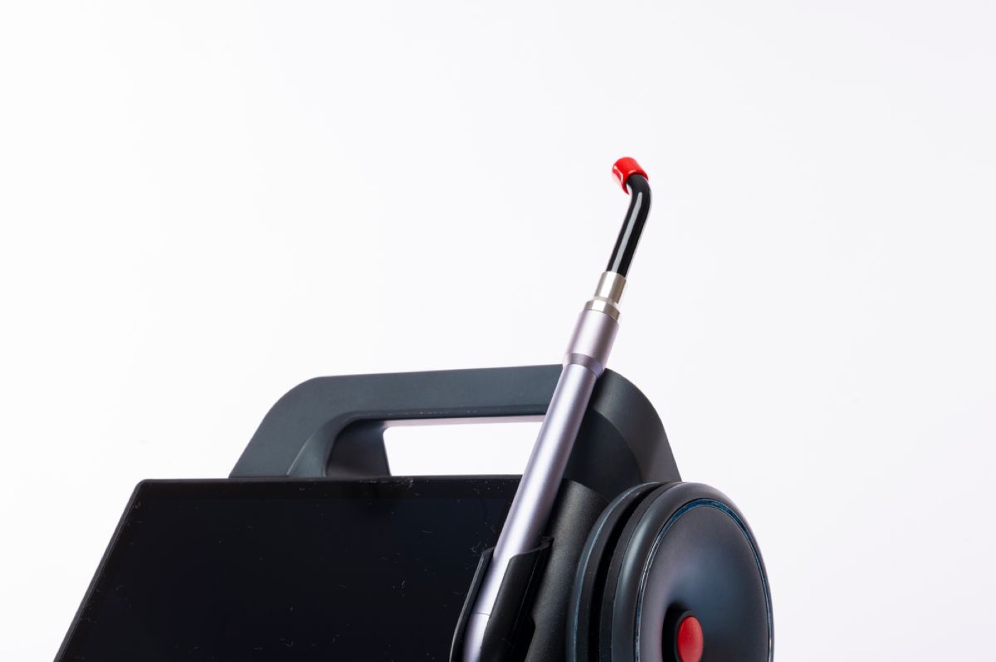
An FDA-cleared laser protocol for treating moderate and advanced gum disease. Its useful property is selectivity — the wavelength targets diseased tissue and bacteria while leaving healthy tissue alone.
Practically, that means we can treat deep gum pockets from the inside using a fibre about the width of three hairs, instead of cutting the gum open and stitching it closed. No scalpel, no sutures, and most patients back to normal the following day.
Digital scanning
Instead of a tray of impression putty held in your mouth while it sets, a small wand photographs your teeth and builds an exact 3D model on screen in a few minutes.
Nothing to gag on, nothing to set, and no repeat appointment when an impression distorts. The digital file goes straight to the laboratory, which is faster and considerably more accurate than a physical mould posted across the country.
Intraoral camera
A small camera that shows you what we're looking at, on a screen, at a size you can actually see.
This one is less about surgery and more about being able to explain things. It's far easier to make a decision about a cracked root or an inflamed pocket when you can see it yourself rather than take our word for it.
PerioView endoscope
A micro-camera on a fibre thin enough to pass below the gumline. It lets us look at a root surface, a fracture or the floor of a pocket directly, instead of working by feel and inference.
Video produced by PerioView, the manufacturer.
Where the laboratory work goes
The crowns, bridges and full arches we fit are made by some of the most reputable dental laboratories in the country. Their technicians come to us — which means for complex cases the person building your teeth can see them in your mouth, match the shade in person, and adjust on the spot rather than by courier.
A note on what technology can't do. None of this substitutes for diagnosis and surgical judgement. A 3D scanner in the hands of someone who doesn't know what they're looking at produces a very detailed picture of a bad plan. The equipment matters because of what our doctors do with it.
Where we use it
Ready to talk it through?
Call any of our three offices, or send us a request and we'll get back to you. Phone hours are Monday to Friday, 7am to 3pm.